Diagram Of Shoulder Joint : Practice, Practice, Practice, Perform: The Shoulder Is Not ...
Diagram Of Shoulder Joint : Practice, Practice, Practice, Perform: The Shoulder Is Not .... It is one of the most mobile joints in the human body, at the cost of joint stability. Antique illustration of human body anatomy: Some signs that you should be seen by a doctor include: This is the smallest rotator cuff muscle. 17 photos of the diagram of shoulder muscles and tendons.
In this episode of eorthopodtv, orthopaedic surgeon randale c. The shoulder joint (glenohumeral joint) is a ball and socket joint between the scapula and the humerus.it is the major joint connecting the upper limb to the trunk. A dislocated shoulder occurs when the humerus (upper arm bone) separates from the shoulder blade at the main shoulder joint. The shoulder joint is composed of the glenoid (the shallow shoulder socket) and the head of the upper arm bone known as the humerus (the ball). A diagram of an anatomic shoulder replacement—the plastic socket replaces the cup of the scapula (shoulder blade).
Distal Clavicle Excision (Resection) - Arlington ... from www.arlingtonortho.com Bursitis (joint inflammation) cervical radiculopathy. It is an extremely mobile joint, in which stability has been sacrificed for mobility. Is the wear and tear of shoulder cartilage until bare bone is exposed. Pronate your wrist so the palm of your hand faces down to the floor (as if you were trying to empty a glass of water). A second joint in the shoulder is the junction of the collar bone with the shoulder blade, called the. Antique illustration of human body anatomy: A metal ball component replaces the worn humeral head. Avascular necrosis (death of bone tissue due to limited blood flow) brachial plexus injury.
While seated or standing, lift the sore arm forward and to the side about thirty to 45 degrees.
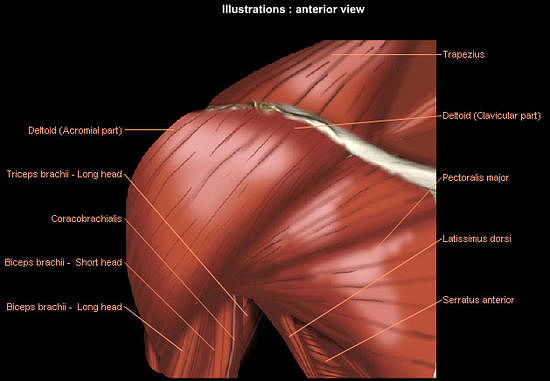
The shoulder joint (glenohumeral joint) is a ball and socket joint between the scapula and the humerus.it is the major joint connecting the upper limb to the trunk. Bones in shoulder, ligaments of the shoulder joint, parts of the shoulder joint, shoulder anatomy, shoulder joints and muscles, shoulder structure anatomy, shoulder tendon anatomy, shoulder tendons ligaments, human muscles, bones in shoulder, ligaments of the shoulder joint, parts of. The shoulder is a complex combination of bones and joints where many muscles act to provide the widest range of motion of any part of the body. The glenohumeral joint is a joint where the greater tubercle (humeral head at the top of the arm bone) meets the shoulder socket of the scapula, called the glenoid cavity or glenoid fossa. 17 photos of the diagram of shoulder muscles and tendons. The shoulder joint is composed of the glenoid (the shallow shoulder socket) and the head of the upper arm bone known as the humerus (the ball). An injury that causes joint deformity. Smartdraw includes 1000s of professional healthcare and anatomy chart templates that you can modify and make your own. The shoulder joint is protected superiorly by an arch, which is formed by the coracoid process of the scapula, the acromion process of the scapula and the clavicle. In this episode of eorthopodtv, orthopaedic surgeon randale c. Diagram of the shoulder, including the location of the rotator cuff. Sechrest, md narrates an animated tutorial on the basic anatomy of the shoulder. Browse 3,854 shoulder anatomy stock photos and images available, or search for shoulder joint or rotator cuff to find more great stock photos and pictures.
Common rotator cuff injuries include rotator cuff tendonitis and rotator cuff strain, which is a partial or complete tear of the rotator cuff. What are common rotator cuff injuries? Ebraheim's educational animated video describes muscle anatomy of the shoulder girdle and anatomy of the shoulder joint.anatomy of the shoulder muscles a. Numerous muscles help stabilize the three joints of. If you have new, worsening, or severe shoulder pain, you should seek medical attention.
Anatomy of Shoulder Joint - PT Master Guide from i1.wp.com Bursitis (joint inflammation) cervical radiculopathy. Smartdraw includes 1000s of professional healthcare and anatomy chart templates that you can modify and make your own. The shoulder has about eight muscles that attach to the scapula, humerus, and clavicle. Bones in shoulder, ligaments of the shoulder joint, parts of the shoulder joint, shoulder anatomy, shoulder joints and muscles, shoulder structure anatomy, shoulder tendon anatomy, shoulder tendons ligaments, human muscles, bones in shoulder, ligaments of the shoulder joint, parts of. The shoulder joint is the junction between the chest and the upper extremity. The shoulder muscles and shoulder tendons involved with shoulder mobility include the four rotator cuff muscle and tendon pairs: The shoulder joint is composed of the glenoid (the shallow shoulder socket) and the head of the upper arm bone known as the humerus (the ball). Browse 3,854 shoulder anatomy stock photos and images available, or search for shoulder joint or rotator cuff to find more great stock photos and pictures.
Its main job is to assist with rotation of the arm away from the body.
Shoulder pain that persists beyond a few days. The shoulder girdle includes three bones—the scapula, clavicle and humerus. Pronate your wrist so the palm of your hand faces down to the floor (as if you were trying to empty a glass of water). Four of them are found on the anterior aspect of the shoulder, whereas the rest are located on the shoulder's posterior aspect and in the back. The shoulder is not a single joint, but a complex arrangement of bones, ligaments, muscles, and tendons that is better called the shoulder girdle. The treatment options are either replacement of just the head of the humerus bone (ball), or replacement of both the ball and the socket (glenoid). Antique illustration of human body anatomy: The shoulder joint can sometimes become narrowed and arthritic, and spurs can form on the undersurface. Some signs that you should be seen by a doctor include: Two joints are at the shoulder. It is one of the most mobile joints in the human body, at the cost of joint stability. There are 10 muscles and 11 shoulder tendons related to shoulder mobility. The top of the humerus is shaped like a ball.
Diagram of the shoulder, including the location of the rotator cuff. Some signs that you should be seen by a doctor include: The muscles in the shoulder aid in a wide. The shoulder is not a single joint, but a complex arrangement of bones, ligaments, muscles, and tendons that is better called the shoulder girdle. Shoulder pain that persists beyond a few days.
Upper body weight training exercises from staticg.sportskeeda.com This diagram depicts muscle diagram of shoulder. The large bone in the upper arm is called the humerus. Its main job is to assist with rotation of the arm away from the body. The shoulder joint is composed of the glenoid (the shallow shoulder socket) and the head of the upper arm bone known as the humerus (the ball). Parts of the right shoulder blade: Some signs that you should be seen by a doctor include: A metal ball component replaces the worn humeral head. The bones of the pectoral girdle (clavicle and scapula) provide increased mobility to the.
The glenohumeral joint is a joint where the greater tubercle (humeral head at the top of the arm bone) meets the shoulder socket of the scapula, called the glenoid cavity or glenoid fossa.
The shoulder is a complex combination of bones and joints where many muscles act to provide the widest range of motion of any part of the body. Two joints are at the shoulder. If you have new, worsening, or severe shoulder pain, you should seek medical attention. Pronate your wrist so the palm of your hand faces down to the floor (as if you were trying to empty a glass of water). What are common rotator cuff injuries? The large bone in the upper arm is called the humerus. Bursitis (joint inflammation) cervical radiculopathy. These muscles form the outer shape of the shoulder and underarm. The components of the ball and cup are reversed on the right—a reverse shoulder replacement. The shoulder girdle includes three bones—the scapula, clavicle and humerus. This diagram depicts muscle diagram of shoulder. Common rotator cuff injuries include rotator cuff tendonitis and rotator cuff strain, which is a partial or complete tear of the rotator cuff. Human anatomy diagrams show internal organs, cells, systems, conditions, symptoms and sickness information and/or tips for healthy living.
This is the smallest rotator cuff muscle diagram of shoulder. Numerous muscles help stabilize the three joints of.


No comments:
Post a Comment